
Dementia is a chronic or progressive syndrome that leads to cognitive decline and fragmentation of memory, thinking, understanding and language. With economic development and the intensification of the aging process, according to data from the World Health Organization (WHO), the number of dementia patients in the world exceeds 55 million, and there are 10 million new confirmed cases each year.
Studies have shown that the brain has undergone functional and structural changes a few years before the onset of cognitive impairment. In addition, metabolic factors such as diabetes, dyslipidemia (high or low blood lipid levels) and hypertension are all related to cognitive decline and dementia. Not only that, patients aged 60 and above with metabolic risk factors are 11.48 times more likely to develop Alzheimer's disease than those without metabolic risk factors.
Therefore, investigating the "relationship between metabolic risk factors and dementia" may help to explore methods for preventing dementia based on improving metabolic risk.
Recently, a study published in the journal Diabetes, Obesity and Metabolism (6.408 points) titled "Metabolic profile-based subgroups can identify differences in brain volumes and brain iron deposition" investigated the relationship between metabolic biomarkers captured by brain imaging data and brain health, which may provide a scientific basis for the prevention of dementia.
The process of this study is clear and very worthy of reference for scientific researchers. Let's take a look!
1. Research background
In fact, the structure and function of the brain have changed long before the symptoms of cognitive impairment are diagnosed. Understanding the metabolic factors and characteristics related to brain changes in the process of dementia will help identify early risk factors for dementia. However, individual metabolic risk factors rarely appear alone, so it is necessary to group the population and assess the risks of related factors caused by gathering together in the population, and then provide prevention methods suitable for individuals.
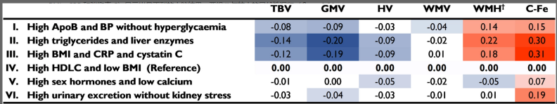
Using a data-driven artificial neural network method, the researchers designed an organizational map (SOM) to detect multivariate patterns in complex data sets. After a metabolic analysis of a large amount of data, the researchers divided the British white population in the UK Biobank population into six subgroups for the assessment of biomarkers related to cardiovascular health, diabetes, kidney and liver function, bones and joints, cancer and obesity. These markers, which can significantly characterize the incidence of diseases, are clustered into subgroups.
In this study, the researchers investigated the relationship between the metabolic subgroups defined by SOM (and their biomarker components) and neuroimaging markers of brain morphology and iron deposition. In addition, the researchers used MRI brain data from the largest survey of its kind (N > 26 000), allowing them to focus on dementia-related measures in the preclinical stage.
2. Research Methods
In this study, the researchers collected 26,239 participants from the UK Biobank who were free of dementia and stroke at baseline. Among them, the age ranged from 37 to 73 years old, and 52% were female. In addition, the researchers also examined the participants' brain MRI data, including:
Total brain volume (TBV)
Gray matter volume (GMV)
White matter hyperintensity (WMH) volume
Hippocampal volume (HV)
Iron deposition
Next, the researchers evaluated brain imaging based on biomarkers, including:
Cholesterol level
Inflammation levels
Kidney function
liver function
Body Mass Index (BMI)
Basal Metabolic Rate (BMR) – the number of calories you burn while resting
Blood pressure
25(OH)D Levels – A Measure of Vitamin D
The most common type of dementia is associated with greater iron deposition in the basal ganglia (including the caudate nucleus, putamen, and globus pallidus).
To estimate the extent of iron deposition in the participants' brains, the researchers selected the caudate nucleus as a representative region (iron deposition in this region has been moderately correlated with measures of cognitive deficits). For the analysis of caudate iron deposition, the researchers converted the median T2* values ​​(in milliseconds) of the left and right caudate nuclei into R2* (R2* = 1/T2*), such that higher R2* values ​​corresponded to greater iron deposition. Individual caudate R2* values ​​were calculated as the average of the left and right caudate R2* values. All brain volume and R2* data were normalized to total head size, and WMH volume data were inversely transformed to approximate a normal distribution.
3. Research results
Among these brain MRI measures, lower total brain volume (TBV) and gray matter volume (GMV), and higher white matter hyperintensities (WMH) volume, were associated with brain damage and functional loss.
Meanwhile, increased iron deposition was associated with an increased risk of Alzheimer's disease. In addition, reduced hippocampal volume (HV) was also associated with poorer memory, language learning, processing speed, and executive function.
Baseline information showed that younger age, higher education, never smoking, longer work hours, and higher levels of physical exercise were associated with higher HV and less brain damage.
They also noted that iron deposition levels increased with age and were higher in smokers and former smokers, those with higher education, longer work hours, and higher levels of vigorous exercise. Among
biomarkers, high triglycerides, high BMI, poor liver and kidney function, and increased inflammation levels were associated with poor brain imaging results.
In particular, high triglycerides and liver dysfunction were associated with the highest levels of iron deposition, brain damage, and GMV loss.
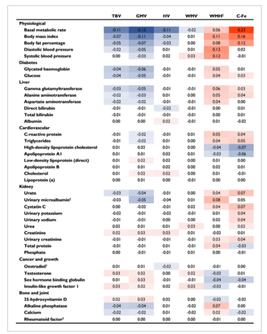
In addition, the researchers noted that a higher basal metabolic rate (BMR) was more associated with more iron deposition, lower GMV, and lower HV than obesity.
They also noted that lower vitamin D levels were associated with more brain damage and lower TBV and GMV.
Overall, there is a significant association between metabolic indicators and brain health. (It is worth noting that although the study found a link between metabolic indicators and brain imaging data of cognitive impairment, it did not resolve causality.)
However, the best way to maintain a healthy metabolism is to avoid excess weight through a healthy diet and physical exercise. Experts suggest that exercise may be the most effective way to prevent Alzheimer's disease!
Source: https://dom-pubs.onlinelibrary.wiley.com/doi/10.1111/dom.14853